Clinical & preclinical research
Custom image analysis for clinical & preclinical research
Turn medical images into quantitative data with analysis and reporting tailored to your needs.
Let’s talk about your imaging workflow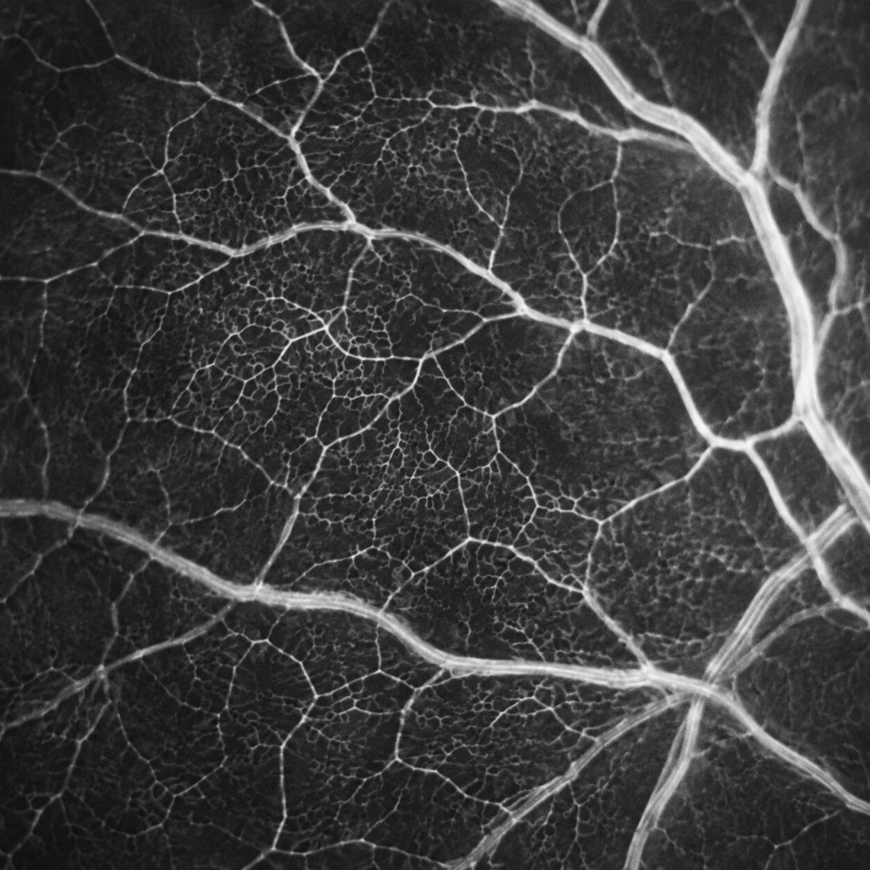
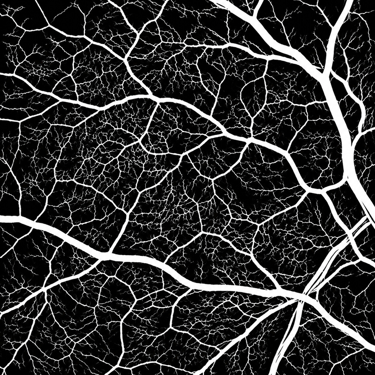
Built around your imaging needs
Different biological and pathology models require different image analysis, measurements and outputs. We customize the analysis and reporting to fit your needs.
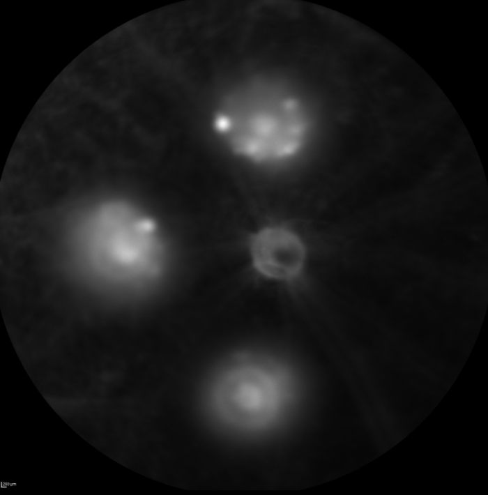
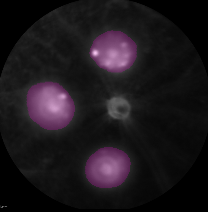
FA image analysis
Segmentation, area, shape, orientation and intensity measurements
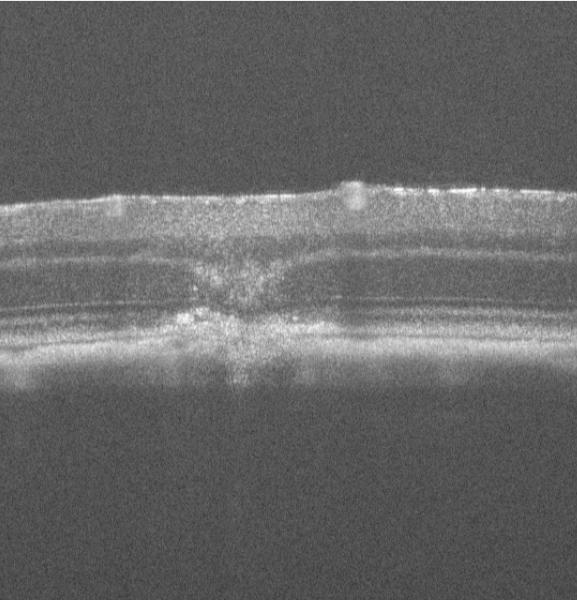
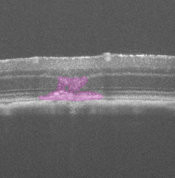
OCT lesion analysis
Segmentation and quantitative measurements
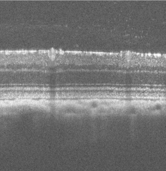
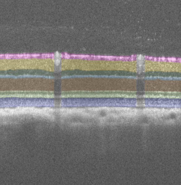
Retinal layer analysis
Multi-layer segmentation and retinal layer thickness
See InVision in action
A short demonstration of the image analysis workflow, from source images to reviewable quantitative results.
Why automate image analysis?
See how repetitive manual measurements can become a faster, reviewable workflow — with less clicking and more time for the science behind the numbers.
Image analysis is its own expertise
Machine learning is only one part. Reliable image analysis also requires geometry, image processing, validation and software engineering.
Complete workflow
From source images to analysis that can be inspected, corrected and used with confidence.
Automated results can be reviewed and corrected. Measurements and reports are then generated from the reviewed analysis.
Have an imaging workflow in mind?
Let’s see what we can do.